Cell culture contaminations: bacteria, yeast, fungi
Sooner or later we all encounter contaminations in our cell cultures. The complicated part is often to identify whether the contamination type is bacteria, yeast or fungi. Knowing what contaminates our cultures might to mitigate the risk for future contamination of our cell culture as the source may be an error in aseptic technique, the water bath, reagents or even the FCS we use.Sometimes cellular debris looks like yeast and sometimes bacteria look like fungi. To help you identify the type of contamination we provide some images that show cells and bacteria, yeast or fungi to give you additional size range and comparison.
Bacteria as cell culture contaminants
Bacteria are by far the most frequent cell culture contamination. This is mostly due to errors in aseptic technique that happen especially when cell culture is performed in a hurry. As bacteria have a very short generation time (minutes to hours) in comparison to mammalian or insect cells, they usually overgrow the cultures very fast (2-3 days) and most often the medium very fast changes to a yellow color. However, especially when antibiotics like penicillin or streptomycin are used in the medium, the initial growth rate might be rather small which makes it harder to identify contamination.
| Strongly contaminated culture with bacterial aggregates | Large bacteria in relation to round yeast | Schematic representation of bacterial morphologies |
 |
 |
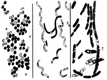 |
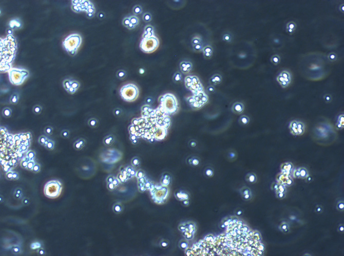
In the image on the left, single bacteria (cocci or rods) and darker bacterial clumps are visible. Kenneth Todar, who supports us on the microbiological side, says that usually cocci have a stronger tendency to form clumps. However, some non-motile rods also form clumps.
|
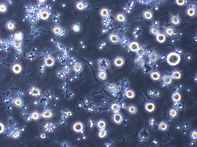
In this image, you see a largely magnified culture of contaminated suspension cells (U937). In comparison, the cells are large and round (largest diameter, yellowish) while the yeast is round but much smaller (white) and the bacteria in this case are attached to one another forming thread-like, dark structures. Usually bacteria appear as small black dots that are even smaller than the bacteria in this image. Even at this magnification it is hard to classify bacteria. Kenneth Todar suggests that the treads are a Bacillus species, maybe Bacillus megaterium. If you want to check on your contamination you might find helpful hints on his page "the normal flora of humans". |
Yeast as a cell culture contamination
Yeast is a much rarer type of contamination found in cell culture. The incidence of yeast usually increases in spring and summer in labs that have a inferior hygiene concept. They are also introduced by errors in asectic technique. Yeast contamination is also easily introduced into insect cell cultures in labs that work with cells and flies. Dry yeast or yeast paste are often added to fly bottles as extra food or to maximise egg laying. It’s important to keep the cell culture and flies totally separate. Yeast cells multiply faster than mammalien cells but slower than bacteria but usually a contamination becomes clearly obvious within 2-3 days in the microscope or it is indicated by the color change of the medium. Yeast are fungi, therefore antibiotics like penicillin and streptomycin have no effect on them.
Yeast cells (400x) in suspension 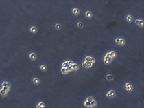 |
Subconfluent HeLa cells with yeast contamination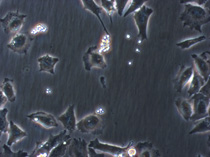 |
Confluent HeLa cells with yeast contamination 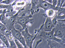 |
|
|
Other fungi as cell culture contaminants
Other fungi are a mean kind of contamination. Usually it is their spores that manage to get into our cultures by an error in aseptic technique through the much easier air-borne route. This means that the spores will take some time to sprout and start to form hyphae, an occurence that we mostly have to find in the microscope by chance. This is the reason why they are often initially overlooked and get the chance to ruin our assays in this way. A safer incubator might help to avoid that.
Large fungal mycelium in a suspension culture (U937) 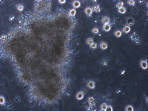 |
Hyphae with two sporangia and smaller spores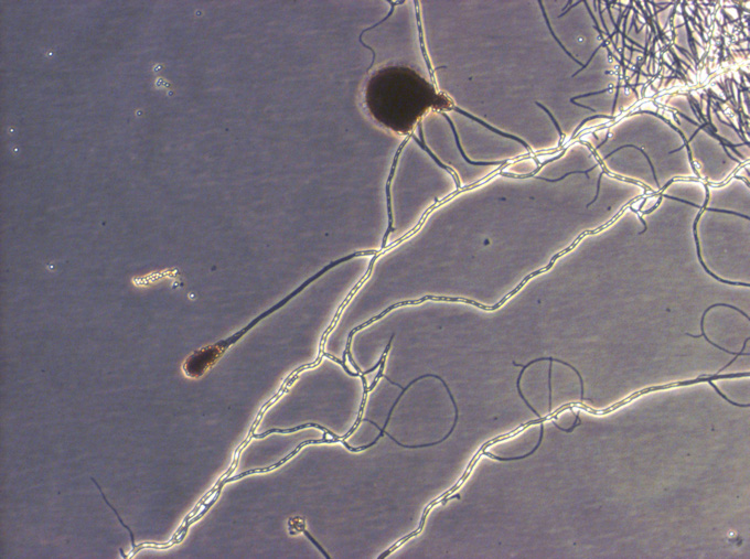 |
Sprouting fungal spores magnified 400x 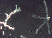 |
|
This is the blown up image from above. In this fungus you can clearly see the separated hyphae (septae) and one large and a smaller sporangium in which fungi produce their spores. On the left you see small, round bright spores that have been released. Note that spores are not killed by ethanol! |
Sources of contamination
In most cases, the reason for contaminations is a mistake in aseptic technique. While newcomers usually make mistakes due to their lack of experience, the growing experience is what endangers long-term cell culturers. The faster we work and the more cultures we handle simuntaneously, the riskier. Below you find a short list of tips that might help you to keep your cultures clean:
- Stress and aseptic tecnique don't go well together
- Always only handle one cell line, cell type in the hood
- Antibiotics cover mistakes. If you can't or don't want to leave them out, try to cultivate a line once in while without antibiotics. This will help you to realize mistakes and additionally, it allows "disguised" contaminations (bacetria supressed by antibiotics, but present) to grow and become visible. This avoids transfer of such contaminations to other cultures.
- Document your contaminations and try to classify them as far as possible. This might help to identify the sourse.
- If you have fungal contaminations once in a while, check your incubator and whether the lab is regularly cleaned with other than alcoholic disinfectants.
- If you have yeast contaminations more often in summer, check your hygiene plan as yeast are usually brought to the lab from outside.
- If you want to be really safe, always test new batches of FCS for bacteria, yeast, fungi and mycoplasma.


